Most moles are harmless, but there are plenty of reasons to want one removed, whether it catches on clothing, affects your confidence or has started to change. Mole removal is a quick, safe procedure under local anaesthetic, but it’s a medical treatment, not a beauty one. Done at home or without a proper assessment, it can lead to infection, avoidable scarring and, most importantly, a missed skin cancer.
Here’s when removal is advisable, how the main techniques work, and what to expect from healing and scarring.
Mole removal falls into two broad groups. The reason for removal shapes which technique is used and what happens to the mole afterwards.
A doctor may recommend removal when a mole shows features that need investigation. These include a mole that:
Doctors often use the ABCDE checklist (Asymmetry, Border, Colour, Diameter, Evolving) as a starting point. The NHS guidance on moles uses similar warning signs. A mole removed for medical reasons is sent to a laboratory for histology, where it is examined under a microscope.
Many people choose mole removal because a mole bothers them. It may be on the face, catch on a razor, bra strap or collar, or affect confidence. Cosmetic removal is entirely reasonable, but only once a doctor is satisfied the mole looks benign. Even then, many doctors still recommend sending the removed tissue for histology as a precaution.
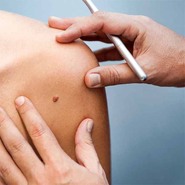
The most important step in any mole removal procedure happens before the procedure itself. A mole that looks harmless to the naked eye can occasionally be an early melanoma or another type of skin cancer.
At your consultation, the doctor will:
If anything about the mole is uncertain, the doctor will recommend a technique that removes it completely and allows full laboratory analysis. If a mole looks suspicious, you may be referred for urgent specialist care rather than cosmetic removal. This is why assessment always comes first: once a mole has been destroyed without a sample, it can no longer be examined.
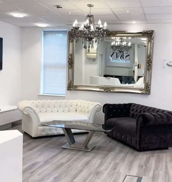
There is no single best way to remove a mole. The doctor chooses the technique based on the mole’s size, depth, location and appearance, as well as your skin type and goals.
Shave excision is often used for raised moles that look benign. After numbing the area, the doctor gently shaves the mole level with the surrounding skin. At Skin Medical, laser may then be used to refine the area for a smoother finish. No stitches are needed, and the area usually heals within one to three weeks. It often leaves a small, flat, slightly paler mark. Because some mole cells may remain below the surface, a small number of moles can partly regrow.
Surgical excision, also called ellipse excision, removes the whole mole, including its deeper root, along with a small margin of surrounding skin. The wound is closed with fine stitches, which are usually removed after 7 to 14 days depending on the area. This method is recommended for flat moles, deeper moles and any mole with uncertain features, as it gives the laboratory a complete sample. It leaves a fine line scar, which is typically longer than the mole itself and fades gradually over time.
Punch excision uses a small circular instrument to remove a small mole in one piece, including its deeper part. One or two stitches may be needed. It suits small moles where a complete sample is needed but a longer ellipse scar is not.
Radiosurgery uses radiofrequency energy to remove or smooth small, raised lesions that have been assessed as benign. It needs no stitches and is often used to refine the edges after shave removal. Cryotherapy, a targeted freezing technique, is generally reserved for skin tags and superficial growths rather than moles.
| Method | Often used for | Stitches | Typical healing | Sample for histology |
|---|---|---|---|---|
| Shave excision (with laser refining) | Raised moles that look benign | No | 1 to 3 weeks | Yes |
| Ellipse (surgical) excision | Flat, deep or uncertain moles | Yes | Stitches out at 7 to 14 days | Yes, complete sample |
| Punch excision | Small moles needing a full-depth sample | Sometimes, 1 or 2 | Stitches out at 7 to 14 days | Yes, complete sample |
| Radiosurgery | Small benign raised lesions, refining edges | No | 1 to 2 weeks | Limited |
Your doctor will explain why a particular method is recommended for you. To see how this works in practice at our Northwood clinic, visit our mole removal clinic page.
Flat mole removal is one of the most searched mole topics, and one where patients are most often given poor advice. A flat mole sits within the skin rather than on top of it. That means it cannot simply be shaved off without leaving a dip or leaving pigment cells behind.
For most flat moles, surgical excision is the recommended approach. It removes the mole completely, allows full histology and gives the doctor control over the direction and length of the scar.
Some services offer to fade flat moles with lasers alone, without a medical assessment first. Laser treatment destroys pigment without producing a sample, so the mole can never be examined. Pigment can also return, and a treated mole that regrows can be harder to assess later. For these reasons, lasers are not suitable for any mole that has not been examined by a doctor.
No. Removing a mole at home is not safe, and neither are unregulated services that skip a medical assessment. Common DIY methods include cutting, tying off with thread, freezing kits and so-called mole removal creams or pens.
These approaches carry real risks:
If you are tempted to remove a mole yourself, a short consultation is a far safer first step.
Any procedure that removes a mole will leave some mark, so mole removal without scarring is not a realistic promise. What a skilled doctor can do is minimise how noticeable that mark is.
This is achieved by choosing the right technique for the mole, placing incisions along the skin’s natural lines, using fine stitches, and giving clear aftercare advice. Scars tend to be more visible on the chest, shoulders and upper back, and some skin types are more prone to raised or darker scars. Your doctor will discuss this with you before treatment.
Most patients go back to work or normal activities the same day. Healing time depends on the method used and where the mole was.
Typical aftercare advice includes:
Scars continue to mature for 12 to 18 months, gradually becoming flatter and paler. Contact the clinic if you notice increasing redness, warmth, swelling, discharge or bleeding that does not stop with gentle pressure.

Mole removal is a common, low-risk procedure, but no procedure is free from risk. Possible side effects include:
It is also worth knowing that removing one mole does not reduce your overall skin cancer risk. Checking your skin regularly and using sun protection remain important.
Skin Medical is an award-winning, doctor-led clinic at the William Old Centre, Ducks Hill Road, Northwood HA6 2NP, with free on-site parking. Patients visit us from across London and nearby areas including Rickmansworth, Pinner, Hatch End, Ruislip, Harrow, Chorleywood and Amersham, with easy access from the M25, M1 and A40.
Our approach to mole removal is built around safety first:
We also remove cysts, lipomas, skin tags and other lesions through our lumps, bumps and skin lesion removal service, and you can explore our full range on our treatments page. For more on why medical oversight matters, read our guide to doctor-led clinics versus high street salons.